Abdomen
Solid Viscera
Describe the kidneys and the variants (30 marks)
DESCRIPTION
- Paired bean-shaped organs that are retroperitoneal.
- Major function is urine production, removing waste products.
- Also has minor endocrine function and is involved with regulation of blood pressure
STRUCTURE
- Anterior and Posterior surface
- Renal hilum as the entrance to renal sinus
- Upper and lower pole
- Medial margin – concave
- Lateral margins – convex
- Fibrous cortex
- Traditionally divided into superior, inferior, two anterior and single posterior “segments” based on its arterial supply
- Renal cortex with renal column (extensions) inward
- Renal pyramids with renal papilla
RENAL HILUM
- Renal hilum is the entrance to space = renal sinus
- Has anterior and posterior lips of renal hilum
- Renal sinus contents:
- Renal pelvis + 2-3x major calyces, supplied by 2-3x minor calyces, supplied by 1x renal papilla (apex of the renal pyramid where urine is excreted)
- Renal artery
- Renal vein
- Nerves
- Fat
- Relations in renal sinus:
- Ureter pelvis exits (most posteriorly!)
- Renal artery enters
- Renal vein exits (most anterior!)
RELATIONS
- Surrounded by perirenal and pararenal fat (see details in abdominal wall and dedicated question)
- Posterior: quadratus lumborum, 12th rib, latissimus dorsi, deep back muscles, thoracolumbar fascia
- Medial: psoas major
- Anterior: peritoneum and abdominal cavity,
- spleen/stomach/pancreas tail/jejunum, descending colon/root of the transverse mesocolon (on left),
- hepatorenal recess, liver, descending duodenum, ascending colon/right colic flexure (on right)
- Lateral: EO, IO, Transverse abdominal muscle
- Superior: weak fascia that separates kidney from adrenal gland, diaphragm, bare area of liver (on right), esophagus (on left)
- Medial: IVC, aorta, celiac plexus, ureters, Sympathetic trunk, T12-L3 vertebral body
- Side note: Perinephric abscess – within the fascia the abscess can only really extend inferiorly between the loosely attached anterior and posterior layers of the pelvic fascia.
ARTERY
- Renal artery – arise at level of IV disc between L1 and L2 vertebrae. Right renal artery is longer and passes posterior to IVC. Each artery divides in 5 segmental arteries at the renal hilum that are end arteries (do not anastomosis)
- Anterior Branch of renal artery
- Superior (apical) segmental renal artery
- *Anterio-superior segmental renal artery
- *Antero-inferior segmental renal artery
- Inferior segmental artery
- Posterior branch of the renal artery
- Posterior segmental artery
- Accessory left renal artery (variant)
- Extrahilar renal arteries (from renal artery of aorta) often enter kidney at the poles (variant)
VEIN
- Right and left renal veins lie anterior to the renal arteries
- Longer left renal vein receives the left adrenal vein, left testicular/ovarian vein and communicates with the ascending lumbar vein, then passes anterior to the aorta. Each renal vein drains into the IVC
LYMPH
- 3x Lymph plexuses of Kidney – caval and aortic lymph nodes = lumbar lymph nodes
- One within kidney parenchyma – 4-5x lymphatic vessels leave renal sinus
- One under the fibrous capsule
- One in the perirenal fat
- 4-5x lymphatic trunks leave the renal sinus and are joined by the lymphatic vessels from the capsule. The lymphatic vessels follow the renal vein to the lumbar (caval and aortic) lymph nodes. Lymph from the suprarenal glands also drains to the lumbar nodes. à chyle cistern
NERVE – aorticorenal plexus
- Renal nerve plexus
- SNS – least splanchnic nerves T11-T12
- Visceral afferent fibres for pain follow sympathetic fibres to T11-L2
VARIANTS (this could be a small question in itself!)
- Extrarenal pelvis (normally pelvis in intrarenal)
- Malrotation of the kidney – increase uretero-pelvic junction obstruction which increases the incidence of urolithiasis and infection, increase the risk of stasis and urolithiasis (hilum normally points anteromedially)
- Nonrotation – hilum faces anteriorly + ureters laterally
- Excess rotation (hyperrotation) – hilum faces posteriorly + renal vessels are posterior
- Reversed rotation – hilum faces laterally + renal vessels anterior + ureter lateral
- Sagittal rotation – kidney rotates around its hilum in the sagittal plane
- Ectopic kidney – ureter has no loose coils or kidneys unlike mobile kidney/nephroptosis
- Accessory left renal artery
- Extrahilar renal arteries (from renal artery of aorta) often enter kidney at the poles – because the kidney develops by ascending to the final site, embryonic kidneys receive their blood supply and venous drainage from successively more superior vessel. The inferior vessel degenerate as superior ones take over the blood supply and venous drainage. Failure of these vessels to degenerate results in accessory renal arteries which enter/exit the poles of the kidney. Occur in 25% people.
- Bifid renal pelvis and ureter – anomalies from division of the ureteric bud (metanephric diverticulum). Extent of the ureteral duplication depends on the completeness of embryonic division of the metanephric diverticulum. The bifid renal pelvis and/or ureter may be unilateral or bilateral; however a separate opening into the bladder are uncommon. Incomplete division of the metanephric diverticulum results in bifid ureter; complete division results in a supernumerary kidney.
- Supernumary kidney = complete division of the metanephric diverticulum (ureteric bud)
- Unilateral duplicated ureter = two ureters into one kidney, but with two openings into the bladder
- Retrocaval ureter – leaves the kidney and passes posterior to the IVC then twists back round
- Horseshoe kidney – inferior poles of right and left kidney fuses – and lies at the L3 vertebrae where the root of the IMA is present and stops it ascending
- Ectopic pelvic kidney – kidney fails to ascend, receives blood from common iliac artery
- Retroaortic left renal vein = “ posterior nutcracker syndrome” – left renal venous hypertension, haematuria, flank pain
- Circumaortic renal vein – left renal vein bifurcates into dorsal and anterior branch to encircle the aorta
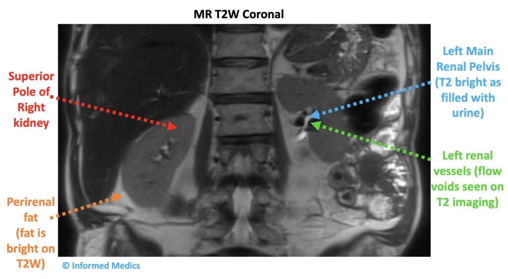

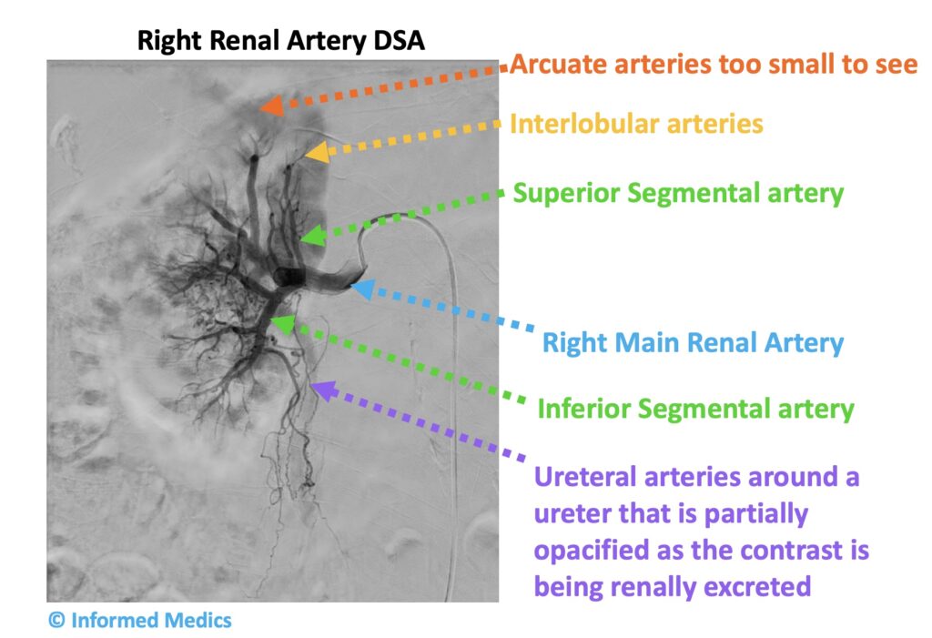
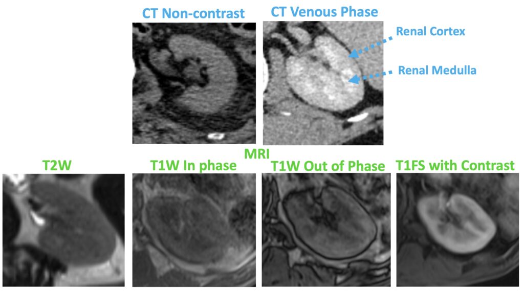
This content is restricted to Anatomy Course Members Please purchase via this link for 12 months access Your account will be activated after payment Please email informedmedics@gmail.com for any questions